 |
Overview |
Loss of vision
poses extraordinary challenges on individuals in our society which relies
heavily on sight. Currently there is no effective treatment for some patients
who are profoundly visually handicapped due to degeneration or damage to the:
New hope has
been generated recently by showing that electrical stimulation of almost any
location along the visual path can evoke the perception of visual perceptions
called “phosphenes”

This
project aims to develop prototypes in the field of visual rehabilitation and to
demonstrate the feasibility of a cortical neuroprosthesis, interfaced with the
visual cortex, as a means through which
a limited but useful visual sense may be restored to profoundly blind people. Even though the full
restoration of vision seems to be impossible, the discrimination of shape and
location of objects could allow blind subjects to ‘navigate’ in a familiar environment and to read
enlarged text, resulting in a substantial improvement in the standard of living of blind
and visually impaired persons.
Several laboratories worldwide are developing microelectronic prosthesis
intended to interact with the remaining healthy retina
or optic nerve.
However the output neurons of the eye (ganglion cell neurons,
which in turn give rise to the optic nerve axons) often degenerate in many
retinal blindnesses and therefore a retinal or optic nerve prosthesis would not
be always helpful. We have decided to work on stimulation of the primary
visual cortex because the neurons in higher visual regions of the brain (visual
cortex) often escape from this degeneration. If these higher visual centers can be
stimulated with visual information in a format somewhat similar to the way they
were stimulated prior to the development of total blindness, a blind individual
may be able to use this stimulation to extract information about the physical
world around him/her. This concept is supported by several studies, which
demonstrate that localized electrical stimulation of the human visual cortex
can excite topographically mapped visual percepts [Bak et al., 1990, Brindley et
al, 1968, 1972; Dobelle et al., 1976, 1978, 2000; Heiduscha and Thanos, 1999;
Normann et al., 1999; Schmidt et al., 1996].

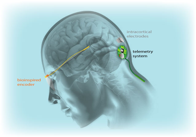
Schema of a Cortical Neuroprosthesis system
The above figure
illustrates the basic components of our approach (Cortical Visual
Neuroprosthesis). The whole system will use a bioinspired (retina-like) visual
processing front-end, able to transform the visual world in front of a blind
individual into electrical signals that can be used to excite, in real time, the neurons at his/her visual cortex. The output
information from this bioinspired peripheral device, will be used to stimulate
an array of penetrating electrodes implanted into the primary visual cortex. Such a visual neuroprosthesis is
expected to recreate a limited, but useful visual sense in a blind individual
who is using such a system.
We are
developing reconfigurable multichannel neurostimulators (128 channels) able to
drive different current injections and waveforms through high impedance
electrodes as well as a multichannel transcutaneous power and data
radiofrequency links to the intracortical microelectrodes. The safety and
efficacy of permanent charge injection through multiple intracortical
electrodes into cerebral cortex is being studied using histopathological and
physiological techniques. We are also using transcranial magnetic
stimulation (TMS), MRI and fMRI to study the degree of remaining functional
visual cortex in blind subjects.
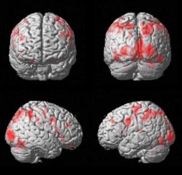
The following functional magnetic resonance imaging (fMRI) study displays the areas of the brain that are activated when a congenital blind person reads Braille letters using our fMRI compatible Braille Stimulator. The areas of activation (displayed in red) are not restricted to the somatosensory cortex cotralateral to the right hand used for reading. Rather, there is marked activation of the occipital cortex, the part of the brain that we use to process visual information.

fMRI of a blind subject reading Braille characteres
We are using these non-invasive techniques, to understand the plastic changes in the brains of persons with visual impairments. Thus, a requirement for clinical application of a cortical prosthesis, aside from safety considerations, is proper, non-invasive patient selection criteria. The primary visual cortex of potential candidates for such neuroprosthetic devices has to be capable of processing visual information, i.e. not have been plastically transformed to process tactile or auditory information. Therefore, the precise understanding and modulation of neuroplastic processes related to the cross-modal transfer of tactile and auditory information to the visual cortex is a critical prerequisite for the implementation of any visual neuroprosthetic approach. In this context one of our goals is to develop a methodology to measure the degree cross-modal plasticity in blind subjects. Another important objective is to develop a methodology to identify the preferred location for the neuroprosthetic device.
The results
from this project will provide information regarding neuroprosthesis not
presently available, as well as new
information about the plastic changes in the adult nervous system, which has
implications for rehabilitative training and device development. If we can
understand more about the fundamental mechanisms of neuronal coding, and to
safely stimulate nervous system, there will real potential to apply this
knowledge clinically. Although we are still a long way from a perfect visual prosthesis,
the success of the cochlear implant seems very promising also for this
neurotechnological application.
References.
Bak M, Girvin JP, Hambrecht FT, Kufta CV, Loeb GE, Schmidt EM. Visual sensation produced by intracortical microstimulation of the human occipital cortex. Med. Biol. Eng. Comput. 28: 257-259 (1990).
Brindley G.S., Lewin W.S. The sensations produced by electrical stimulation of the visual cortex. J. Physiol. 196:479-493 (1968).
Brindley GS, Donaldson PEK, Falconer MA, Rushton DN. The extent of the region of occipital cortex that when stimulated gives phosphenes fixed in the visual field. J. Physiol (Lond) 225: 57P-58P (1972).
Dobelle WH, Mladejovsky MG, Evans JR, Roberts TS, Girvin JP. 'Braille' reading by a blind volunteer by visual cortex stimulation. Nature, 259: 111-112(1976).
Dobelle WH, Mladejovsky MG, Girvin JP. Artificial vision for the blind: electrical stimulation of visual cortex offers hope for a functional prothesis. Science, 183: 440-444 (1974).
Dobelle WH, Mladejowsky MG. Phosphenes produced by electrical stimulation of human occipital cortex, and their application to the development of a prothesis for the blind. J. Physiol (Lond) 243: 553-576(1974).
Dobelle, W.H. Artificial vision for the blind by connecting a television camera to the visual cortex. ASAIO Journal 2000; 46: 3-9.
Heiduschka P, Thanos S. Implantable bioelectronic interfaces for lost nerve functions. Prog. Neurobiol. 55: 433-461 (1998).
Normann RA, Maynard, E., Rousche, PJ, Warren DJ. A neural interface for a cortical vision prosthesis. Vision Res 1999 Jul;39(15):2577-87
Schmidt EM, Bak MJ, Hambrecht FT, Kufta CV, O'Rourke DKO, Vallabhanath P. Feasibility of a visual prothesis for the blind based on intracortical microstimulation of the visual cortex. Brain, 119: 507-522 (1996).